Home

persuadere Hobart lago wga staining protocol richiedente A bordo Esci

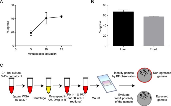
A fast, non-invasive, quantitative staining protocol provides insights in  Plasmodium falciparum gamete egress and in the role of osmiophilic bodies |  Malaria Journal | Full Text
A fast, non-invasive, quantitative staining protocol provides insights in Plasmodium falciparum gamete egress and in the role of osmiophilic bodies | Malaria Journal | Full Text

Wheat germ agglutinin (WGA) staining pattern in myocardial cells before...  | Download Scientific Diagram
Wheat germ agglutinin (WGA) staining pattern in myocardial cells before... | Download Scientific Diagram
Wheat Germ Agglutinin (WGA) Conjugates | ATLANTIS BIOSCIENCE

Wheat Germ Agglutinin Wga (Vector Laboratories) | Bioz | Ratings For  Life-Science Research
Wheat Germ Agglutinin Wga (Vector Laboratories) | Bioz | Ratings For Life-Science Research

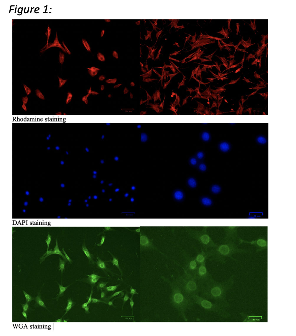
Alvetex Protocol: Wheat Germ Agglutinin Staining

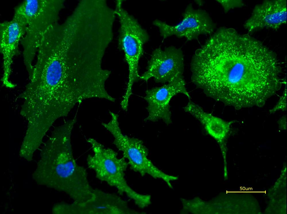
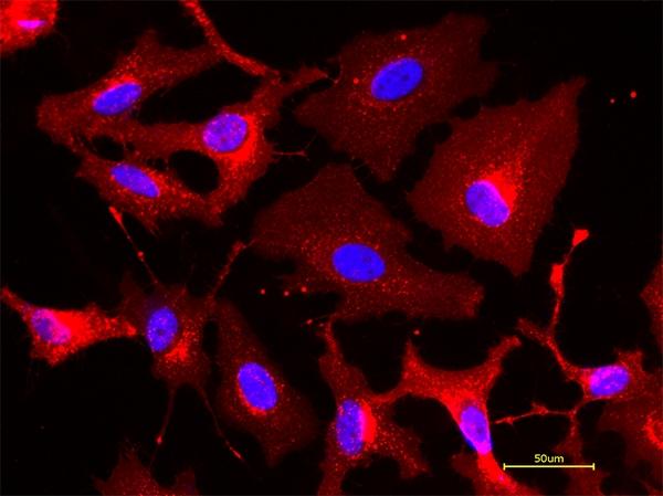
iFluor® 488-Wheat Germ Agglutinin (WGA) Conjugate | AAT Bioquest

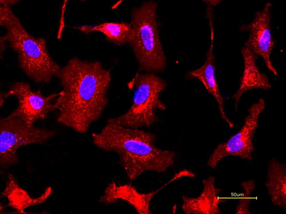
iFluor® 647-Wheat Germ Agglutinin (WGA) Conjugate | AAT Bioquest

Membrane & Cell Surface Stains

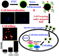
Wheat germ agglutinin modified magnetic iron oxide nanocomplex as a cell  membrane specific receptor target material for killing breast cancer cells  - Journal of Materials Chemistry B (RSC Publishing)
Wheat germ agglutinin modified magnetic iron oxide nanocomplex as a cell membrane specific receptor target material for killing breast cancer cells - Journal of Materials Chemistry B (RSC Publishing)

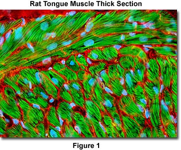
Confocal Microscopy - Tissue Cryosection Triple Stain | Olympus LS

Triple immunohistochemical staining for the visualization of fibrous... |  Download Scientific Diagram
Triple immunohistochemical staining for the visualization of fibrous... | Download Scientific Diagram

PDF] Wheat Germ Agglutinin Staining as a Suitable Method for Detection and  Quantification of Fibrosis in Cardiac Tissue after Myocardial Infarction |  Semantic Scholar
PDF] Wheat Germ Agglutinin Staining as a Suitable Method for Detection and Quantification of Fibrosis in Cardiac Tissue after Myocardial Infarction | Semantic Scholar
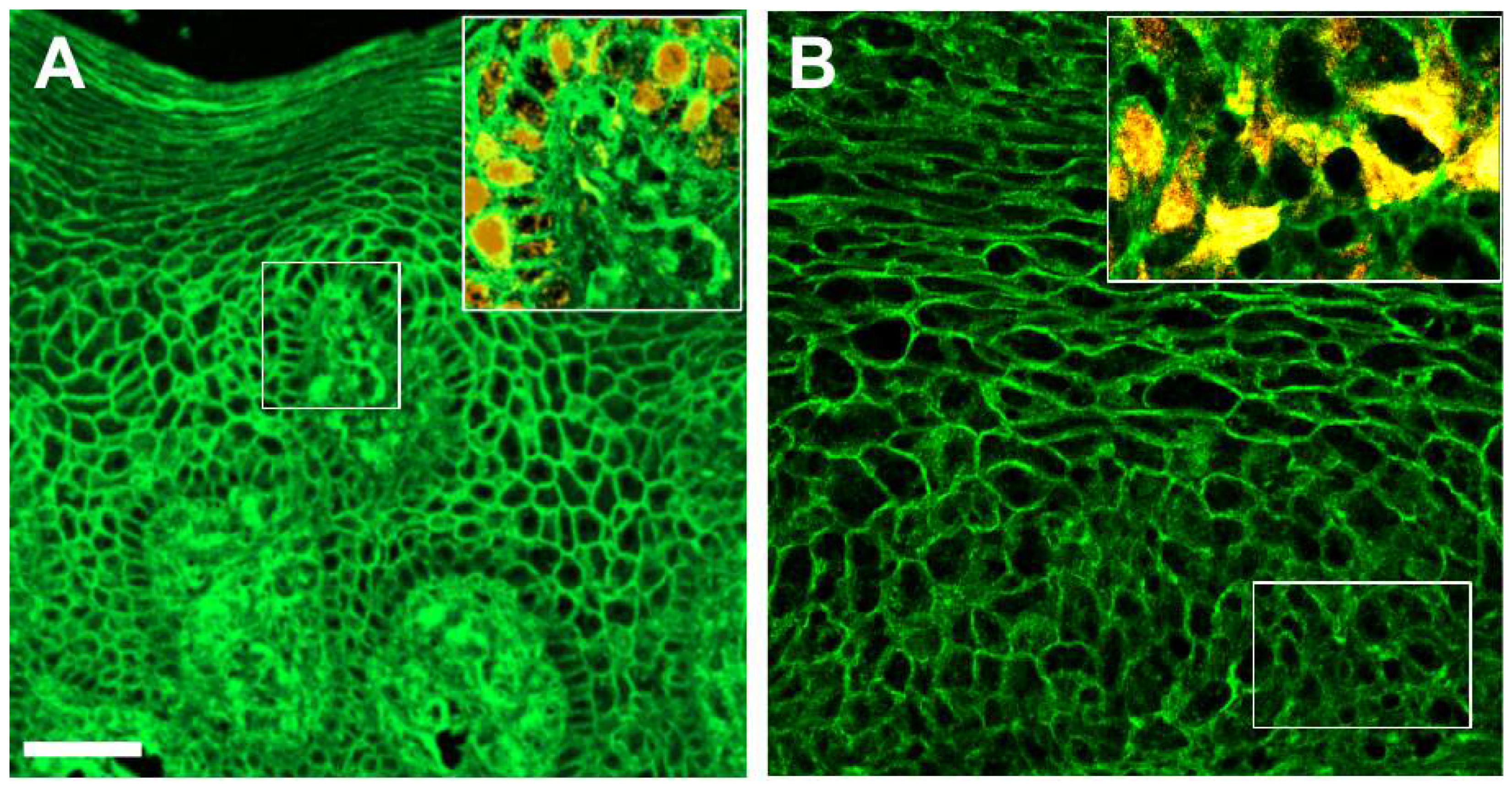
Cells | Free Full-Text | The Tissue Architecture of Oral Squamous Cell  Carcinoma Visualized by Staining Patterns of Wheat Germ Agglutinin and  Structural Proteins Using Confocal Microscopy
Cells | Free Full-Text | The Tissue Architecture of Oral Squamous Cell Carcinoma Visualized by Staining Patterns of Wheat Germ Agglutinin and Structural Proteins Using Confocal Microscopy
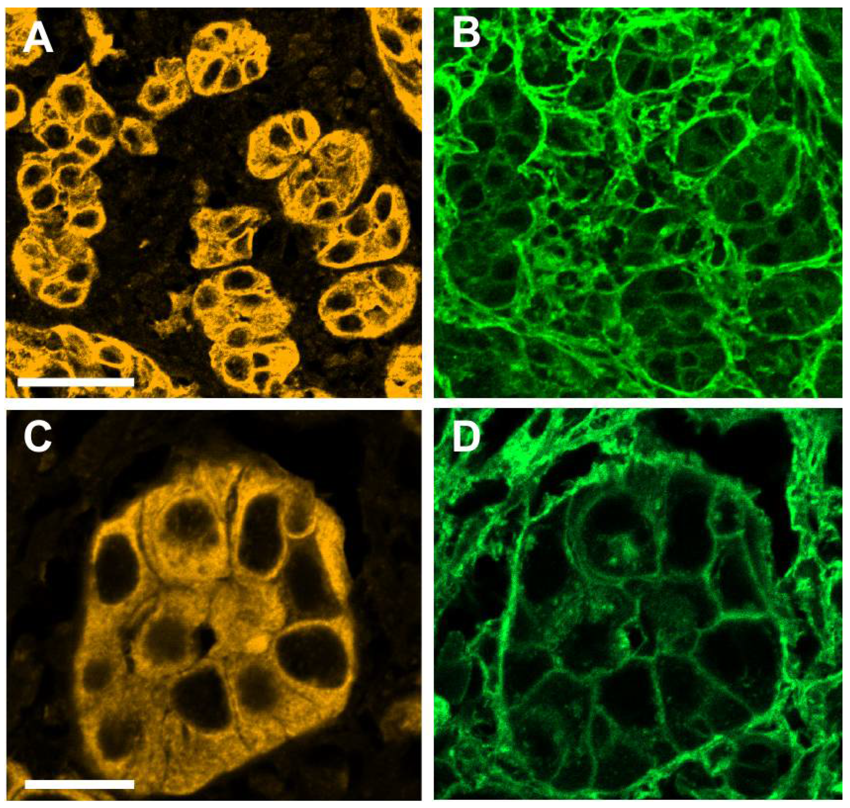

Wheat Germ Agglutinin (WGA) Conjugates | ATLANTIS BIOSCIENCE

iFluor® 594-Wheat Germ Agglutinin (WGA) Conjugate | AAT Bioquest

An optimized protocol for combined fluorescent lectin/immunohistochemistry  to characterize tissue-specific glycan distribution in human or rodent  tissues - ScienceDirect
An optimized protocol for combined fluorescent lectin/immunohistochemistry to characterize tissue-specific glycan distribution in human or rodent tissues - ScienceDirect

PDF] Wheat Germ Agglutinin Staining as a Suitable Method for Detection and  Quantification of Fibrosis in Cardiac Tissue after Myocardial Infarction |  Semantic Scholar
PDF] Wheat Germ Agglutinin Staining as a Suitable Method for Detection and Quantification of Fibrosis in Cardiac Tissue after Myocardial Infarction | Semantic Scholar

Cells | Free Full-Text | The Tissue Architecture of Oral Squamous Cell  Carcinoma Visualized by Staining Patterns of Wheat Germ Agglutinin and  Structural Proteins Using Confocal Microscopy
Cells | Free Full-Text | The Tissue Architecture of Oral Squamous Cell Carcinoma Visualized by Staining Patterns of Wheat Germ Agglutinin and Structural Proteins Using Confocal Microscopy

CellBrite® Fix Membrane Stains - Biotium

Validation and flow chart of the wheat germ agglutinin-staining... |  Download Scientific Diagram
Validation and flow chart of the wheat germ agglutinin-staining... | Download Scientific Diagram

PhenoVue Fluor - Wheat Germ Agglutinin Conjugates

Solved For the Cellular Localization and Fluorescence | Chegg.com

Three-dimensional direct measurement of cardiomyocyte volume, nuclearity,  and ploidy in thick histological sections
Three-dimensional direct measurement of cardiomyocyte volume, nuclearity, and ploidy in thick histological sections