Home

Danimarca cultura sgattaiolare nissl staining protocol tintura pazzo cane

Nissl stain sections corresponding to MRI slices. Nissl stained... |  Download Scientific Diagram
Nissl stain sections corresponding to MRI slices. Nissl stained... | Download Scientific Diagram

Does anyone have a working protocol for cresyl violet preparation for nissl  staininig? | ResearchGate
Does anyone have a working protocol for cresyl violet preparation for nissl staininig? | ResearchGate

Nissl body - Wikipedia

Protocol to assess the effect of disease-driving variants on mouse brain  morphology and primary hippocampal neurons - ScienceDirect
Protocol to assess the effect of disease-driving variants on mouse brain morphology and primary hippocampal neurons - ScienceDirect

Does anyone have a working protocol for cresyl violet preparation for nissl  staininig? | ResearchGate
Does anyone have a working protocol for cresyl violet preparation for nissl staininig? | ResearchGate
Nissl staining - YouTube

Staining neurons - YouTube

Histological methods for CNS

Chemical Probes for Visualizing Intact Animal and Human Brain Tissue

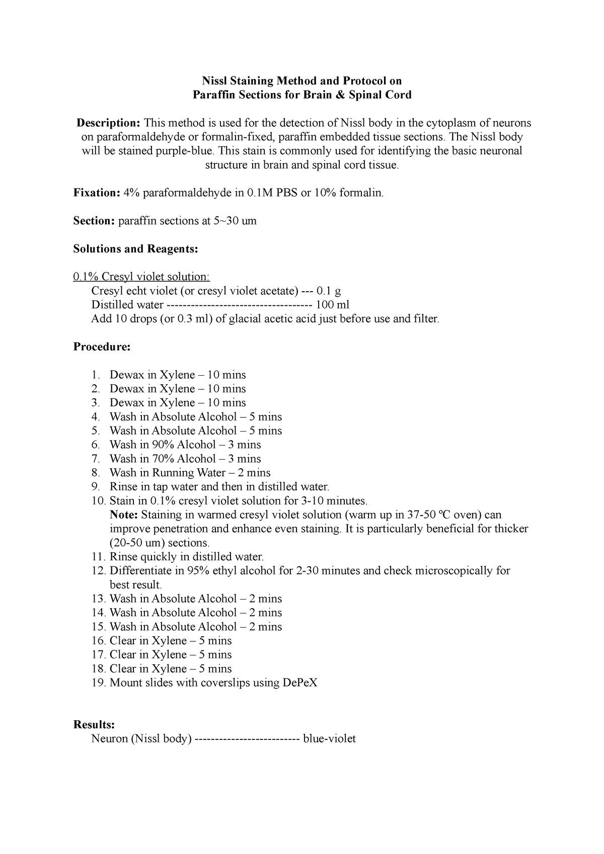
Nissl Staining Method brain - The Nissl body will be stained purple-blue.  This stain is commonly - Studocu
Nissl Staining Method brain - The Nissl body will be stained purple-blue. This stain is commonly - Studocu

Nissl Staining - an overview | ScienceDirect Topics

Combined histochemical staining, RNA amplification, regional, and single  cell cDNA analysis within the hippocampus | Laboratory Investigation
Combined histochemical staining, RNA amplification, regional, and single cell cDNA analysis within the hippocampus | Laboratory Investigation

Histological methods for CNS

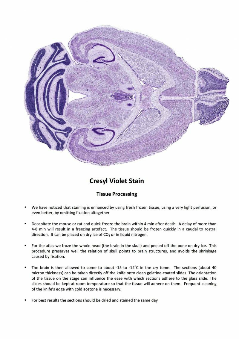
Cresyl Violet Staining (Nissl Staining)

The fate of Nissl-stained dark neurons following traumatic brain injury in  rats: difference between neocortex and hippocampus regarding survival rate  | SpringerLink
The fate of Nissl-stained dark neurons following traumatic brain injury in rats: difference between neocortex and hippocampus regarding survival rate | SpringerLink

Representative sections of the Nissl staining of different brain... |  Download Scientific Diagram
Representative sections of the Nissl staining of different brain... | Download Scientific Diagram

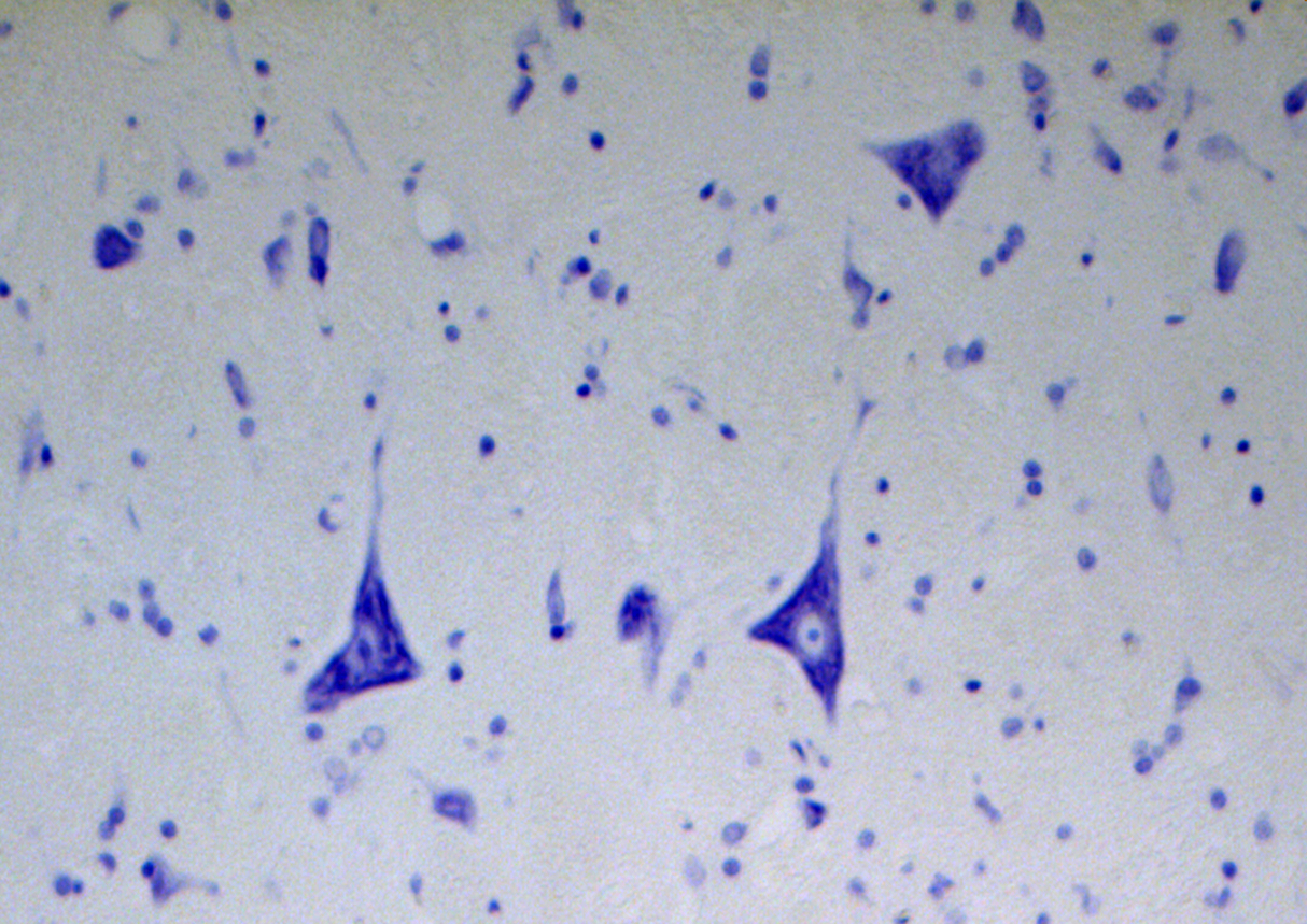
What is a Nissl stain? — Brain Stuff

Nissl Body - an overview | ScienceDirect Topics

Nissl stain analysis. A: After Nissl staining, the typical morphology... |  Download Scientific Diagram
Nissl stain analysis. A: After Nissl staining, the typical morphology... | Download Scientific Diagram
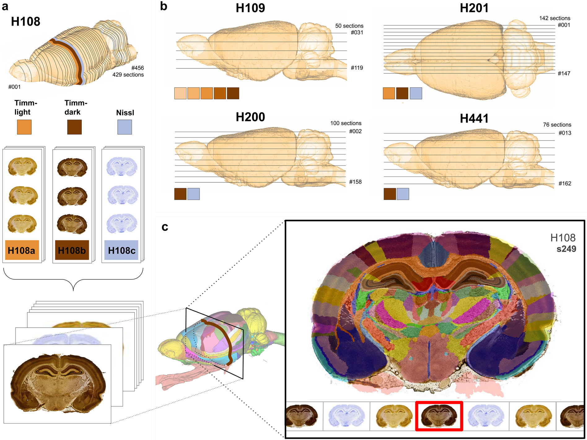
A Timm-Nissl multiplane microscopic atlas of rat brain zincergic terminal  fields and metal-containing glia | Scientific Data
A Timm-Nissl multiplane microscopic atlas of rat brain zincergic terminal fields and metal-containing glia | Scientific Data

stains of nervous tissues.pptx

VitroView™ Nissl Stain Kit - Detection of Nissl body in the cytoplasm of  neurons
VitroView™ Nissl Stain Kit - Detection of Nissl body in the cytoplasm of neurons

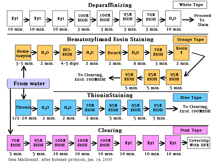
Rubelab
Rubelab

NeuRA | George Paxinos | Neuroscience Research Australia

Nissl body identification in DRG tissue using Nissl staining method (n... |  Download Scientific Diagram
Nissl body identification in DRG tissue using Nissl staining method (n... | Download Scientific Diagram

Nissl staining - دید سبز Nissl staining % Nissl staining

Electroacupuncture Increases the Expression of Gas7 and NGF in the  Prefrontal Cortex of Male Rats with Focal Cerebral Ischemia
Electroacupuncture Increases the Expression of Gas7 and NGF in the Prefrontal Cortex of Male Rats with Focal Cerebral Ischemia

Representative images of Nissl staining in the hippocampus (A), dentate...  | Download Scientific Diagram
Representative images of Nissl staining in the hippocampus (A), dentate... | Download Scientific Diagram