Home

Mittens acquistare Archeologia lvot vti echo musica lirica merge tramonto

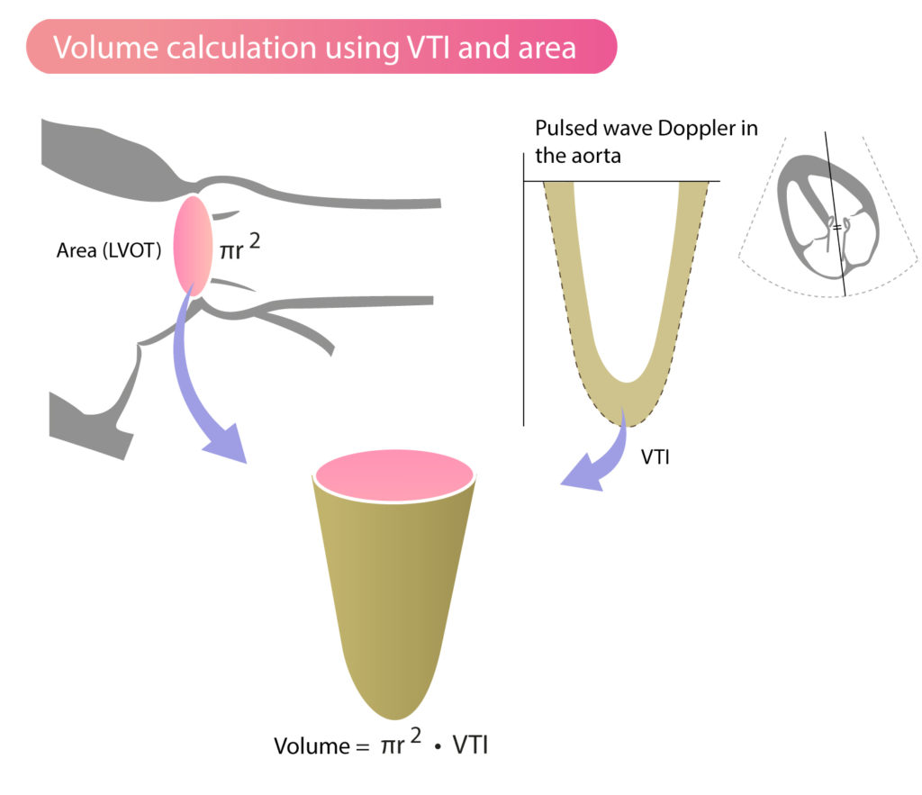
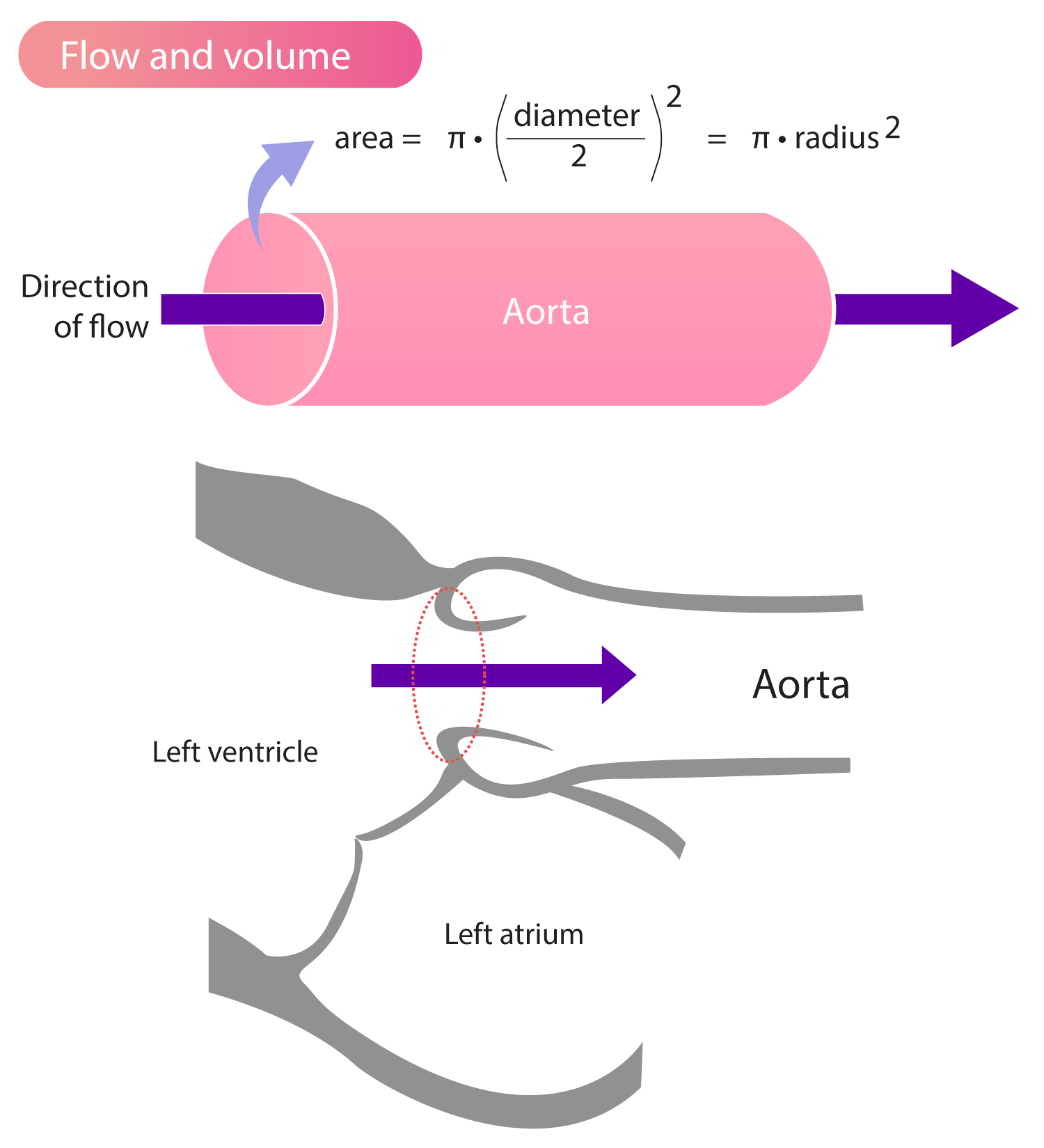
How To Calculate Stroke Volume In Echocardiography (The EASY Way)

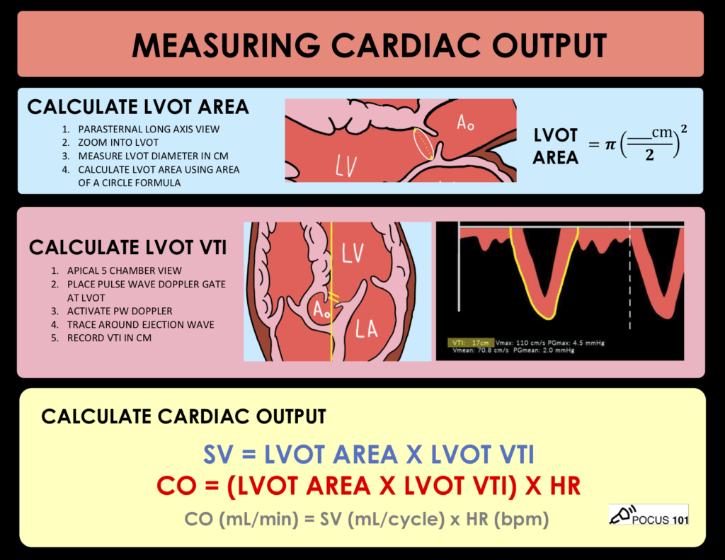
Measuring Cardiac Output with Echocardiography Made Easy - POCUS 101

Accurate stroke volume (SV) estimation: SV = LVOT area × LVOT VTI. a... |  Download Scientific Diagram
Accurate stroke volume (SV) estimation: SV = LVOT area × LVOT VTI. a... | Download Scientific Diagram

A, Normal LVOT VTI (VTI TSVI, 19.09 cm), indicating a normal stroke... |  Download Scientific Diagram
A, Normal LVOT VTI (VTI TSVI, 19.09 cm), indicating a normal stroke... | Download Scientific Diagram

Left ventricular outflow tract velocity-time integral: A proper measurement  technique is mandatory - Pablo Blanco, 2020
Left ventricular outflow tract velocity-time integral: A proper measurement technique is mandatory - Pablo Blanco, 2020

Stroke Volume, VTI (Velocity Time Integral) & Cardiac Output – ECG & ECHO

Stroke Volume, VTI (Velocity Time Integral) & Cardiac Output – ECG & ECHO

Automation of sub-aortic velocity time integral measurements by  transthoracic echocardiography: clinical evaluation of an artificial  intelligence-enabled tool in critically ill patients - British Journal of  Anaesthesia
Automation of sub-aortic velocity time integral measurements by transthoracic echocardiography: clinical evaluation of an artificial intelligence-enabled tool in critically ill patients - British Journal of Anaesthesia

Left ventricular outflow tract velocity time integral in hospitalized heart  failure with preserved ejection fraction - Omote - 2020 - ESC Heart Failure  - Wiley Online Library
Left ventricular outflow tract velocity time integral in hospitalized heart failure with preserved ejection fraction - Omote - 2020 - ESC Heart Failure - Wiley Online Library

Smart VTI
Smart VTI

Measuring Cardiac Output with Echocardiography Made Easy - POCUS 101

Cardiac Output
Cardiac Output

Absolute values of the left ventricular tract velocity-time integral... |  Download Scientific Diagram
Absolute values of the left ventricular tract velocity-time integral... | Download Scientific Diagram
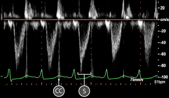
Virtual TEE: Using spectral Doppler to evaluate the left ventricular  outflow tract
Virtual TEE: Using spectral Doppler to evaluate the left ventricular outflow tract

Normal Blood Flow Patterns

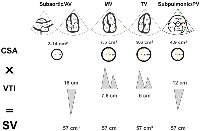
Rationale for using the velocity–time integral and the minute distance for  assessing the stroke volume and cardiac output in point-of-care settings |  The Ultrasound Journal | Full Text
Rationale for using the velocity–time integral and the minute distance for assessing the stroke volume and cardiac output in point-of-care settings | The Ultrasound Journal | Full Text

Left ventricular outflow tract velocity time integral outperforms ejection  fraction and Doppler-derived cardiac output for predicting outcomes in a  select advanced heart failure cohort | Cardiovascular Ultrasound | Full Text
Left ventricular outflow tract velocity time integral outperforms ejection fraction and Doppler-derived cardiac output for predicting outcomes in a select advanced heart failure cohort | Cardiovascular Ultrasound | Full Text

Velocity Time Integral (VTI): Clinical Tips for Your Patients in Shock –  CriticalCareNow
Velocity Time Integral (VTI): Clinical Tips for Your Patients in Shock – CriticalCareNow

NephroPOCUS on X: "@BJegorovic Cardiac output calculation using echo.  Normal VTI is ~18-22 cm. This patient had ~26. https://t.co/Y12pzm2Lk0" / X
NephroPOCUS on X: "@BJegorovic Cardiac output calculation using echo. Normal VTI is ~18-22 cm. This patient had ~26. https://t.co/Y12pzm2Lk0" / X
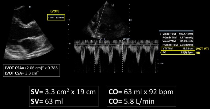
Rationale for using the velocity–time integral and the minute distance for  assessing the stroke volume and cardiac output in point-of-care settings |  The Ultrasound Journal | Full Text
Rationale for using the velocity–time integral and the minute distance for assessing the stroke volume and cardiac output in point-of-care settings | The Ultrasound Journal | Full Text

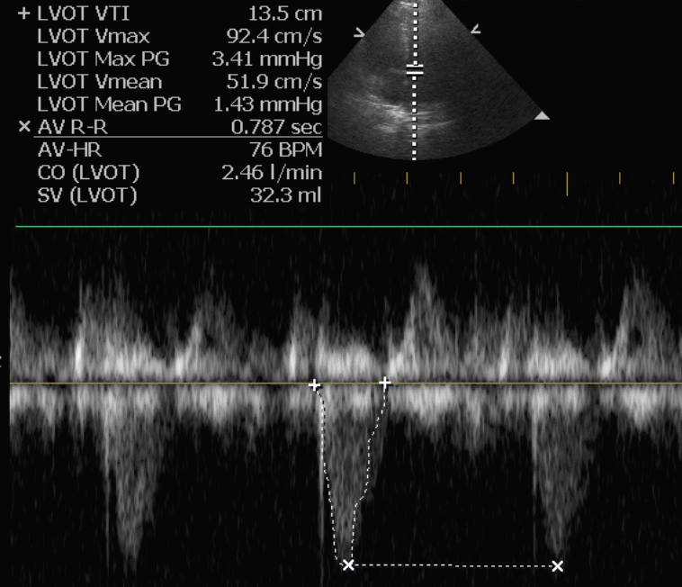
Example image of pulse wave Doppler in the LVOT and measurement of VTI... |  Download Scientific Diagram
Example image of pulse wave Doppler in the LVOT and measurement of VTI... | Download Scientific Diagram
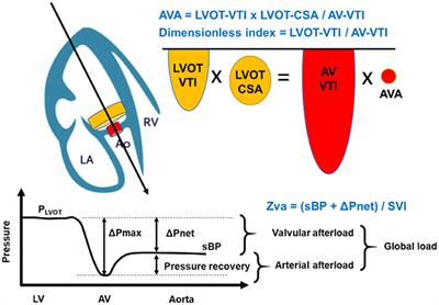
Frontiers | Valvulo-Arterial Impedance and Dimensionless Index for Risk  Stratifying Patients With Severe Aortic Stenosis
Frontiers | Valvulo-Arterial Impedance and Dimensionless Index for Risk Stratifying Patients With Severe Aortic Stenosis

Point of Care Echo: Stroke Volume Determination - YouTube

Aortic Stenosis: Breaking Down the Continuity Equation Cardioserv

a Left ventricular outflow tract velocity-time integral (LVOT VTI) in... |  Download Scientific Diagram
a Left ventricular outflow tract velocity-time integral (LVOT VTI) in... | Download Scientific Diagram