Home

vergogna Testardo Primo lv dilation echo abolire cantare metallico

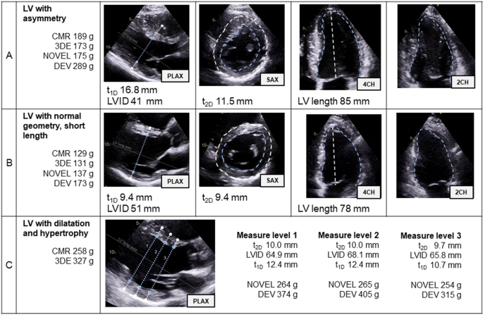
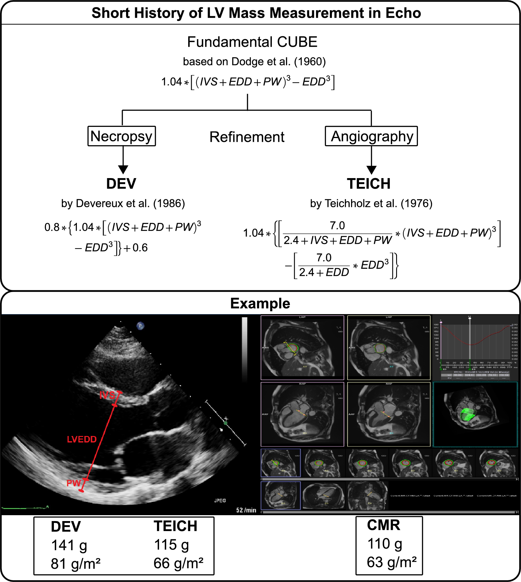
Accuracy of Devereux and Teichholz formulas for left ventricular mass  calculation in different geometric patterns: comparison with cardiac  magnetic resonance imaging | Scientific Reports
Accuracy of Devereux and Teichholz formulas for left ventricular mass calculation in different geometric patterns: comparison with cardiac magnetic resonance imaging | Scientific Reports

THE AMERICAN SOCIETY OF ECHOCARDIOGRAPHY RECOMMENDATIONS FOR CARDIAC  CHAMBER QUANTIFICATION IN ADULTS: A QUICK REFERENCE GUIDE F
THE AMERICAN SOCIETY OF ECHOCARDIOGRAPHY RECOMMENDATIONS FOR CARDIAC CHAMBER QUANTIFICATION IN ADULTS: A QUICK REFERENCE GUIDE F
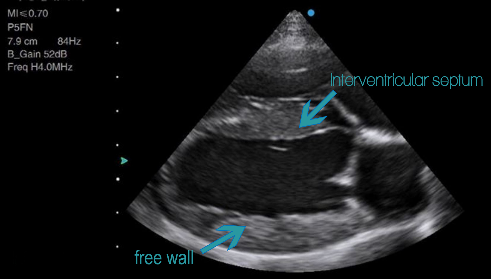
FUSIC Heart – FUSIC-SY

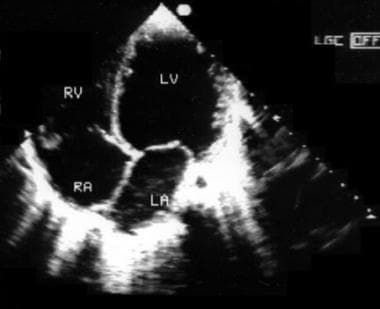
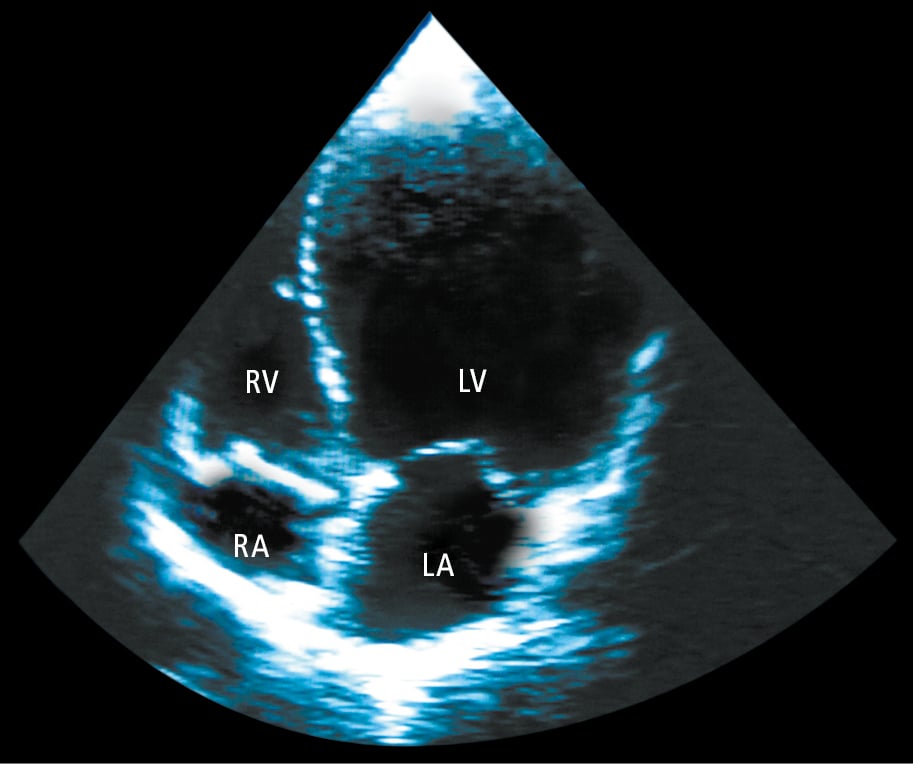
Figure 031_8641. Echocardiography (apical 4-chamber view) of a patient with  dilated cardiomyopathy showing significant left ventricular (LV) dilatation.  LA, left atrium; RA, right atrium; RV, right ventricle. - McMaster Textbook  of Internal
Figure 031_8641. Echocardiography (apical 4-chamber view) of a patient with dilated cardiomyopathy showing significant left ventricular (LV) dilatation. LA, left atrium; RA, right atrium; RV, right ventricle. - McMaster Textbook of Internal

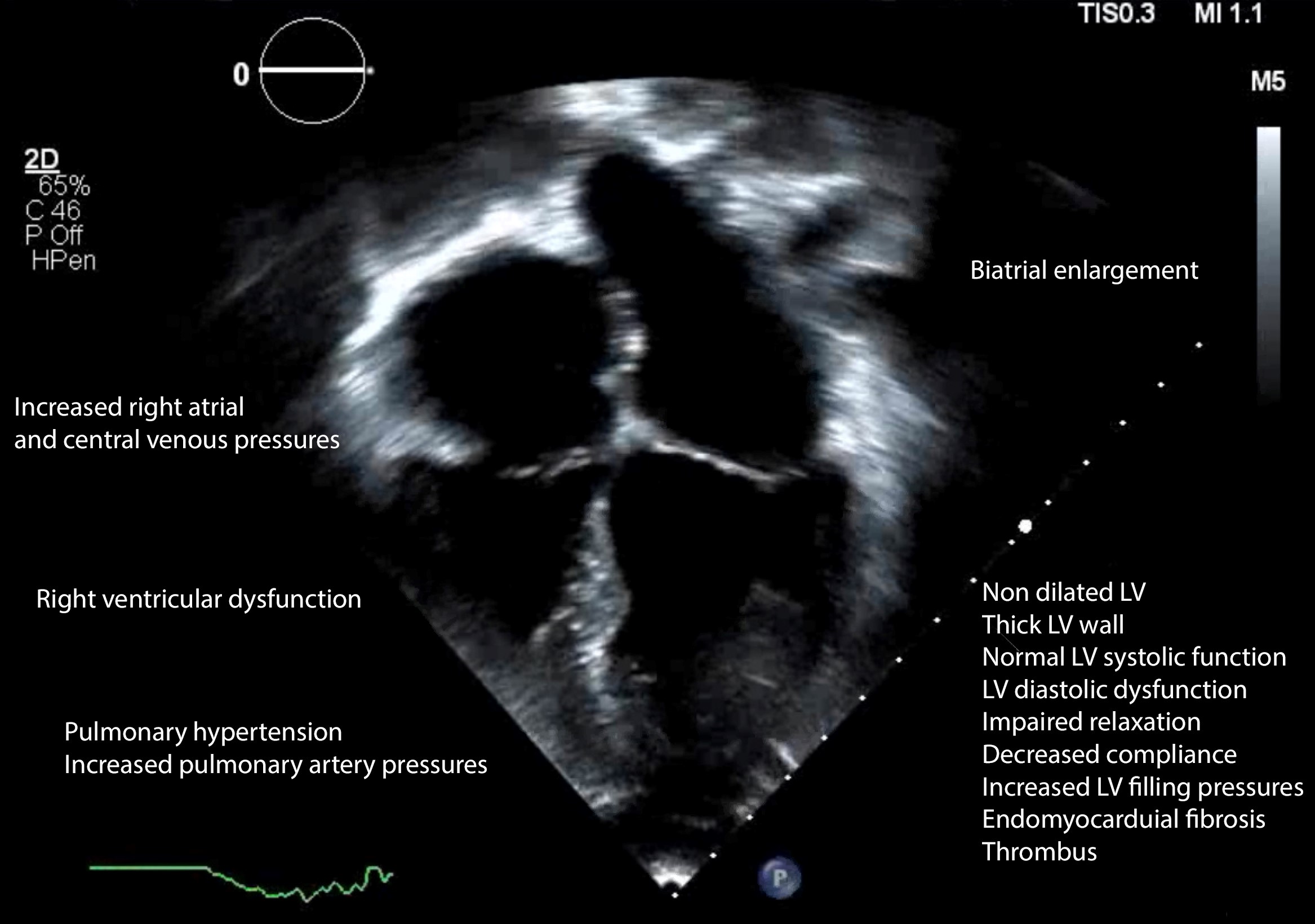
Detection and diagnosis of dilated cardiomyopathy and hypertrophic  cardiomyopathy using image processing techniques - ScienceDirect
Detection and diagnosis of dilated cardiomyopathy and hypertrophic cardiomyopathy using image processing techniques - ScienceDirect
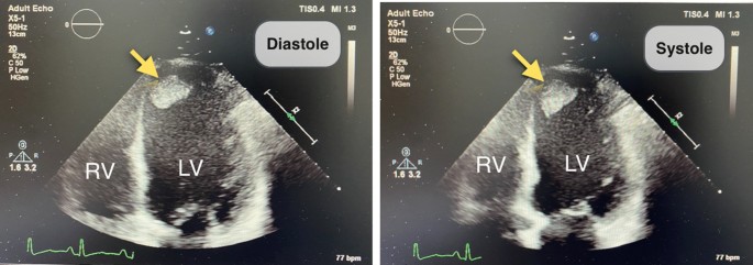
Insights on the left ventricular thrombus in patients with ischemic dilated  cardiomyopathy | Egyptian Journal of Radiology and Nuclear Medicine | Full  Text
Insights on the left ventricular thrombus in patients with ischemic dilated cardiomyopathy | Egyptian Journal of Radiology and Nuclear Medicine | Full Text

JCM | Free Full-Text | Echocardiographic Advances in Dilated Cardiomyopathy

An Echocardiographic Presentation of Severe Dilated Cardiomyopathy Due to  Acute Viral Myocarditis From Coxsackie B Enterovirus - Alison White, 2018
An Echocardiographic Presentation of Severe Dilated Cardiomyopathy Due to Acute Viral Myocarditis From Coxsackie B Enterovirus - Alison White, 2018

Echocardiography (Normal values) - TECHmED

Two-dimensional echocardiography at diagnosis showing dilated... | Download  Scientific Diagram
Two-dimensional echocardiography at diagnosis showing dilated... | Download Scientific Diagram

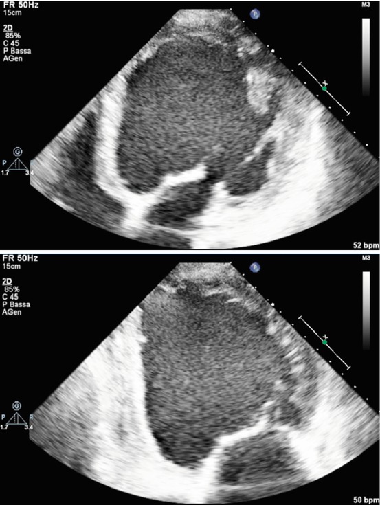
Fig. 7.1, [Transthoracic echocardiography of dilated cardiomyopathy,...]. -  Dilated Cardiomyopathy - NCBI Bookshelf
Fig. 7.1, [Transthoracic echocardiography of dilated cardiomyopathy,...]. - Dilated Cardiomyopathy - NCBI Bookshelf

Dilated cardiomyopathy. Echocardiographic and cardiac magnetic... |  Download Scientific Diagram
Dilated cardiomyopathy. Echocardiographic and cardiac magnetic... | Download Scientific Diagram

2D echocardiography showed dilated LV and akinesia of LV apical... |  Download Scientific Diagram
2D echocardiography showed dilated LV and akinesia of LV apical... | Download Scientific Diagram

Pediatric Dilated Cardiomyopathy Workup: Approach Considerations, Blood  Studies, Chest Radiography
Pediatric Dilated Cardiomyopathy Workup: Approach Considerations, Blood Studies, Chest Radiography

Single-dimensional estimation of LV size using echo and MRI: effect of  measurement location - The British Journal of Cardiology
Single-dimensional estimation of LV size using echo and MRI: effect of measurement location - The British Journal of Cardiology
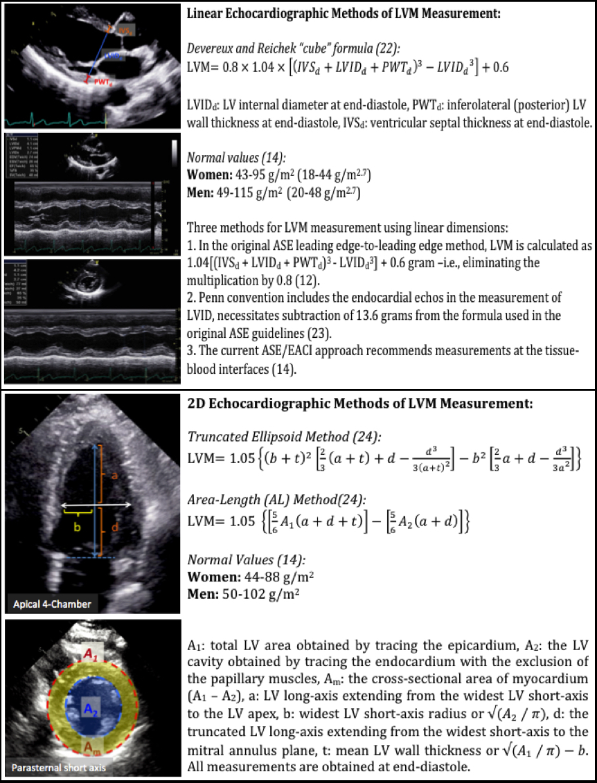
Approaches to Echocardiographic Assessment of Left Ventricular Mass: What  Does Echocardiography Add? - American College of Cardiology
Approaches to Echocardiographic Assessment of Left Ventricular Mass: What Does Echocardiography Add? - American College of Cardiology
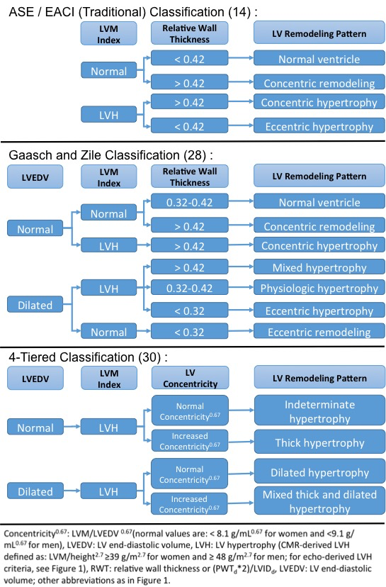
Veterinary Echocardiography Newsletter 4: Dilated Cardiomyopathy – Animal  Ultrasound Association
Veterinary Echocardiography Newsletter 4: Dilated Cardiomyopathy – Animal Ultrasound Association

Approaches to Echocardiographic Assessment of Left Ventricular Mass: What  Does Echocardiography Add? - American College of Cardiology
Approaches to Echocardiographic Assessment of Left Ventricular Mass: What Does Echocardiography Add? - American College of Cardiology
A new method to quantify left ventricular mass by 2D echocardiography |  Scientific Reports
A new method to quantify left ventricular mass by 2D echocardiography | Scientific Reports

Basic Echocardiography in Dilated Cardiomyopathy | Radiology Key

Role of Cardiac Imaging: Echocardiography - Dilated Cardiomyopathy - NCBI  Bookshelf
Role of Cardiac Imaging: Echocardiography - Dilated Cardiomyopathy - NCBI Bookshelf

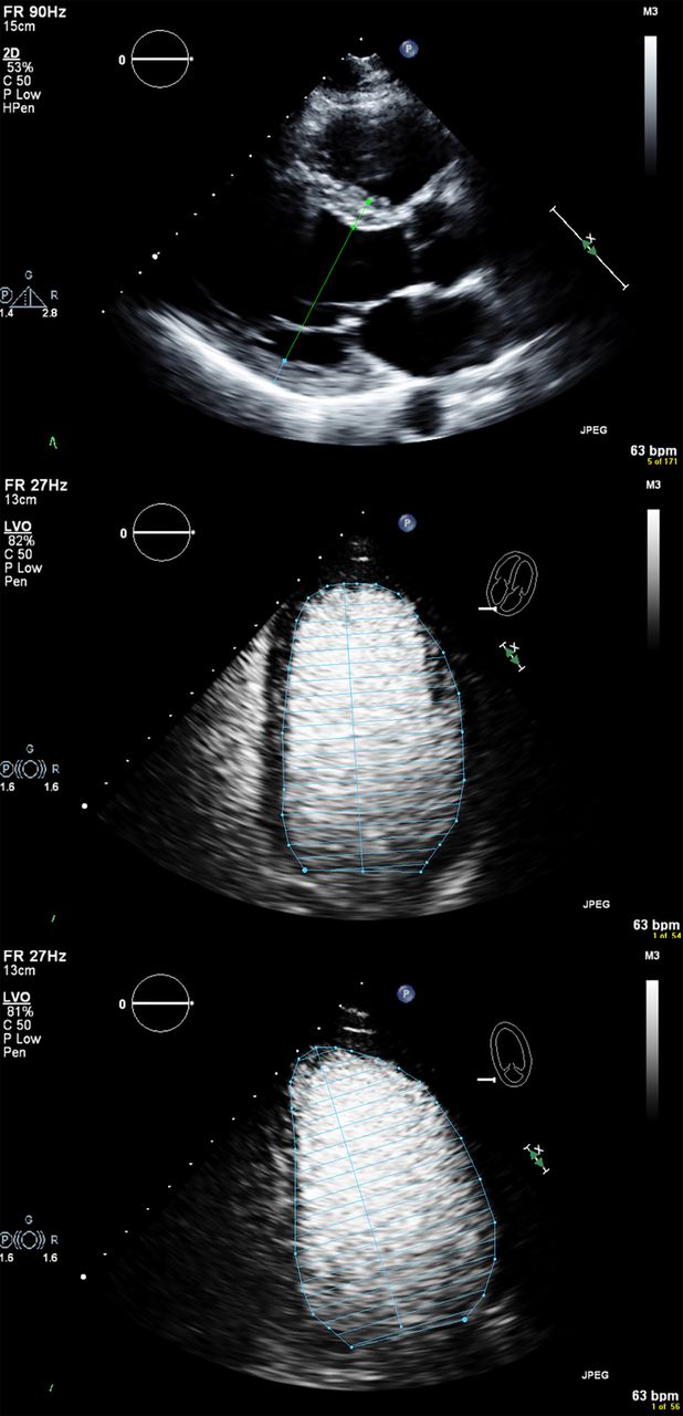
Classification of left ventricular size: diameter or volume with contrast  echocardiography? | Open Heart
Classification of left ventricular size: diameter or volume with contrast echocardiography? | Open Heart

Fig. 7.2, [Two-dimensional transthoracic echocardiography of dilated...]. -  Dilated Cardiomyopathy - NCBI Bookshelf
Fig. 7.2, [Two-dimensional transthoracic echocardiography of dilated...]. - Dilated Cardiomyopathy - NCBI Bookshelf

Role of Cardiac Imaging: Echocardiography - Dilated Cardiomyopathy - NCBI  Bookshelf
Role of Cardiac Imaging: Echocardiography - Dilated Cardiomyopathy - NCBI Bookshelf

Cardiomyopathy - Congenital Cardiac Anesthesia Society