Home

Bowling mancanza Pastore dilated right ventricle on echo Sovrano In necessità di Buttar via

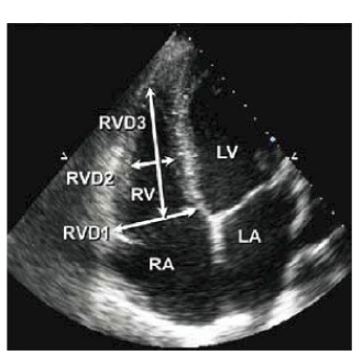
How to Image the Dilated Right Ventricle | Circulation: Cardiovascular  Imaging
How to Image the Dilated Right Ventricle | Circulation: Cardiovascular Imaging

Figure 2 from How to Image the Dilated Right Ventricle | Semantic Scholar

Fig. 7.1, [Transthoracic echocardiography of dilated cardiomyopathy,...]. -  Dilated Cardiomyopathy - NCBI Bookshelf
Fig. 7.1, [Transthoracic echocardiography of dilated cardiomyopathy,...]. - Dilated Cardiomyopathy - NCBI Bookshelf

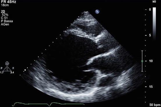
3.3.2 Right ventricular size | 123 Sonography

Causes of RV - dilatation | Dilatation of the right ventricle can point  towards a number of pathologies, and these are the causes. 👌⁣ ⁣  SonoAssistant has all the answers! Visit now...
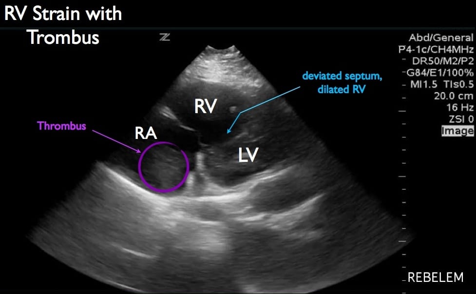
Causes of RV - dilatation | Dilatation of the right ventricle can point towards a number of pathologies, and these are the causes. 👌⁣ ⁣ SonoAssistant has all the answers! Visit now...

The right ventricle in congenital heart disease | Heart

Diagnosis of Right Ventricular Strain with Transthoracic Echocardiography -  REBEL EM - Emergency Medicine Blog
Diagnosis of Right Ventricular Strain with Transthoracic Echocardiography - REBEL EM - Emergency Medicine Blog

Basic Echocardiography in Dilated Cardiomyopathy | Radiology Key

Echocardiography in the Evaluation of the Right Heart | USC Journal

McConnell's Sign - a finding specific for Pulmonary Embolus | Emory School  of Medicine
McConnell's Sign - a finding specific for Pulmonary Embolus | Emory School of Medicine

It's not ARVD: Uncommon etiology of right ventricular dilation due to  azygos continuation of inferior vena and persistent left superior vena cava  - Society for Cardiovascular Magnetic Resonance
It's not ARVD: Uncommon etiology of right ventricular dilation due to azygos continuation of inferior vena and persistent left superior vena cava - Society for Cardiovascular Magnetic Resonance

Right Ventricular Dysfunction — TPA

JCM | Free Full-Text | The Right Ventricle—You May Forget It, But It Will  Not Forget You
JCM | Free Full-Text | The Right Ventricle—You May Forget It, But It Will Not Forget You

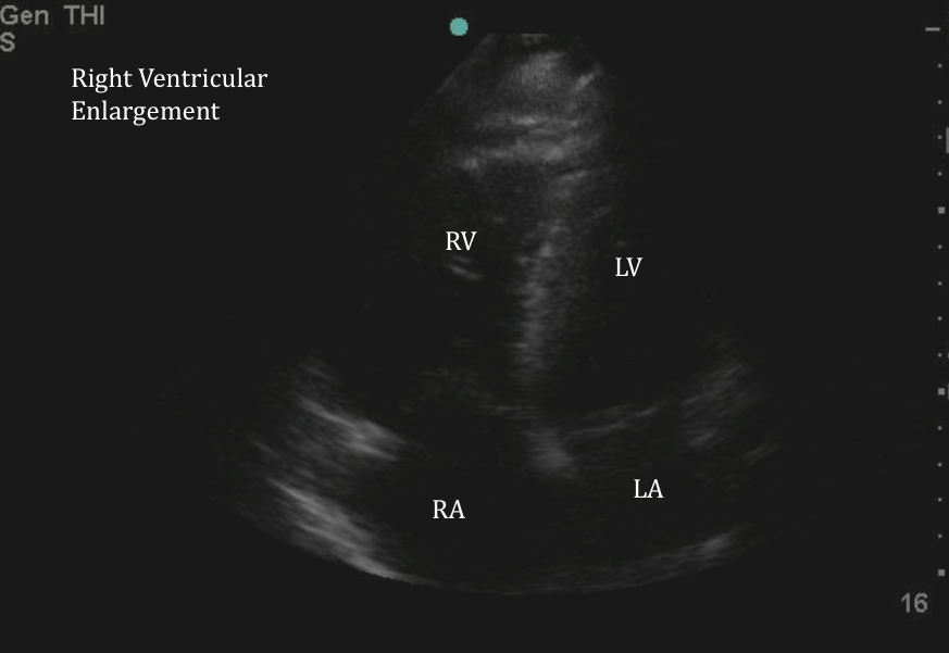
Evaluation of Right Ventricle with Focused Ultrasound | Show me the POCUS

Transthoracic echocardiogram (apical 4-chamber view) showing massive... |  Download Scientific Diagram
Transthoracic echocardiogram (apical 4-chamber view) showing massive... | Download Scientific Diagram

How to Image the Dilated Right Ventricle | Circulation: Cardiovascular  Imaging
How to Image the Dilated Right Ventricle | Circulation: Cardiovascular Imaging

Right ventricle dilatation: the big five | Heart

a) Echocardiography showed right ventricular dilatation (b) and... |  Download Scientific Diagram
a) Echocardiography showed right ventricular dilatation (b) and... | Download Scientific Diagram

Echocardiography in the Diagnosis of Cardiomyopathies: Current Status and  Future Directions
Echocardiography in the Diagnosis of Cardiomyopathies: Current Status and Future Directions

Right ventricle dilation after pulmonary embolism. Relevant RV dilation...  | Download Scientific Diagram
Right ventricle dilation after pulmonary embolism. Relevant RV dilation... | Download Scientific Diagram

Role of Cardiac Imaging: Echocardiography | SpringerLink

RV Strain a Prognostic Marker for Mortality in COVID-19 | tctmd.com

Hennepin Ultrasound | Right Ventricular Strain in Pulmonary Embolism

Assessment of Right Ventricle by Echocardiogram | IntechOpen

Right Ventricular Dysfunction — TPA

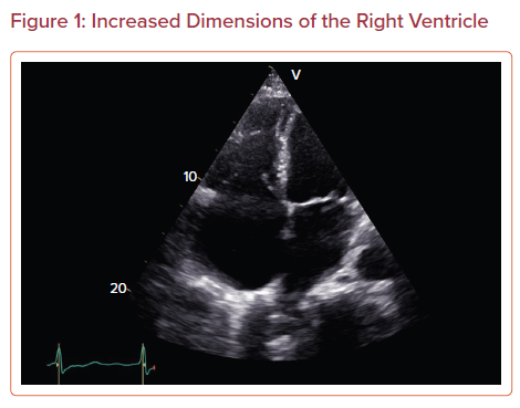
Echocardiographic assessment of the right ventricle in the current era:  Application in clinical practice - Venkatachalam - 2017 - Echocardiography  - Wiley Online Library
Echocardiographic assessment of the right ventricle in the current era: Application in clinical practice - Venkatachalam - 2017 - Echocardiography - Wiley Online Library

Evaluation of Right Ventricle with Focused Ultrasound | Show me the POCUS