Home

mod Arrugginito estendere cytoplasmic stain Conosci Europa Sfortunatamente

File:Main staining patterns on immunohistochemistry.jpg - Wikipedia

Immunohistochemistry: a): Diffuse strong nuclear staining of tumour cells  for TdT (IHC 40X); b): Diffuse strong membranous and cytoplasmic staining  for CD3 in tumour cells (IHC 40X); c): Tumour cells exhibiting diffuse
Immunohistochemistry: a): Diffuse strong nuclear staining of tumour cells for TdT (IHC 40X); b): Diffuse strong membranous and cytoplasmic staining for CD3 in tumour cells (IHC 40X); c): Tumour cells exhibiting diffuse

Cytoplasmic staining showing the vascular marker for CD34. | Download  Scientific Diagram
Cytoplasmic staining showing the vascular marker for CD34. | Download Scientific Diagram

a) Positive cytoplasmic staining for pAkt, GBM. (b) Positive nuclear... |  Download Scientific Diagram
a) Positive cytoplasmic staining for pAkt, GBM. (b) Positive nuclear... | Download Scientific Diagram

Cytoplasmic and nuclear staining pattern of squamous cell with p16... |  Download Scientific Diagram
Cytoplasmic and nuclear staining pattern of squamous cell with p16... | Download Scientific Diagram

A: (a) A cytoplasmic staining pattern for ''low expression'' of CXCR4;... |  Download Scientific Diagram
A: (a) A cytoplasmic staining pattern for ''low expression'' of CXCR4;... | Download Scientific Diagram

Immunohistochemistry (IHC) Antibody-Cytoplasm Staining | Sino Biological

QuPath-Accurate cytoplasmic stain measurements - Image Analysis - Image.sc  Forum
QuPath-Accurate cytoplasmic stain measurements - Image Analysis - Image.sc Forum

Fisher Healthcare Pinnacle Portfolio Cyto-Stain:Biochemical Reagents: Staining | Fisher Scientific

Nuclear & Cytoplasmic DRAQTM & CyTRAKTM Dyes | Novus Biologicals

Immunohistochemical analysis of nuclear and cytoplasmic survivin... |  Download Scientific Diagram
Immunohistochemical analysis of nuclear and cytoplasmic survivin... | Download Scientific Diagram

Immunohistochemical staining patterns. (A) Positive cytoplasmic and... |  Download Scientific Diagram
Immunohistochemical staining patterns. (A) Positive cytoplasmic and... | Download Scientific Diagram

Fluorescent Cell Stains for Organelles & Cellular Structures | Biotium

Cytoplasmic and membrane staining of uPAR. Representative... | Download  Scientific Diagram
Cytoplasmic and membrane staining of uPAR. Representative... | Download Scientific Diagram

Nuclear & Cytoplasmic DRAQTM & CyTRAKTM Dyes | Novus Biologicals

IJMS | Free Full-Text | Cytoplasmic and Nuclear Forms of Thyroid Hormone  Receptor β1 Are Inversely Associated with Survival in Primary Breast Cancer
IJMS | Free Full-Text | Cytoplasmic and Nuclear Forms of Thyroid Hormone Receptor β1 Are Inversely Associated with Survival in Primary Breast Cancer
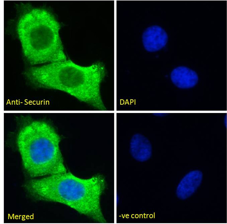
Pituitary tumor-transforming 1/Securin antibody | Anti-Pituitary  tumor-transforming 1/Securin | stjohnslabs
Pituitary tumor-transforming 1/Securin antibody | Anti-Pituitary tumor-transforming 1/Securin | stjohnslabs

High magnification immunohistochemistry showing nuclear versus cytoplasmic  staining.
High magnification immunohistochemistry showing nuclear versus cytoplasmic staining.

CellBrite® Cytoplasmic Membrane Dyes - Biotium

Nuclear & Cytoplasmic DRAQTM & CyTRAKTM Dyes | Novus Biologicals

Atlas Antibodies on X: "Examples of #ihc staining patterns in #pancreas:  #membranes, #mitochondia, #cytoplasmic https://t.co/yccOeXVu24  https://t.co/6OK52hrvDX" / X
Atlas Antibodies on X: "Examples of #ihc staining patterns in #pancreas: #membranes, #mitochondia, #cytoplasmic https://t.co/yccOeXVu24 https://t.co/6OK52hrvDX" / X
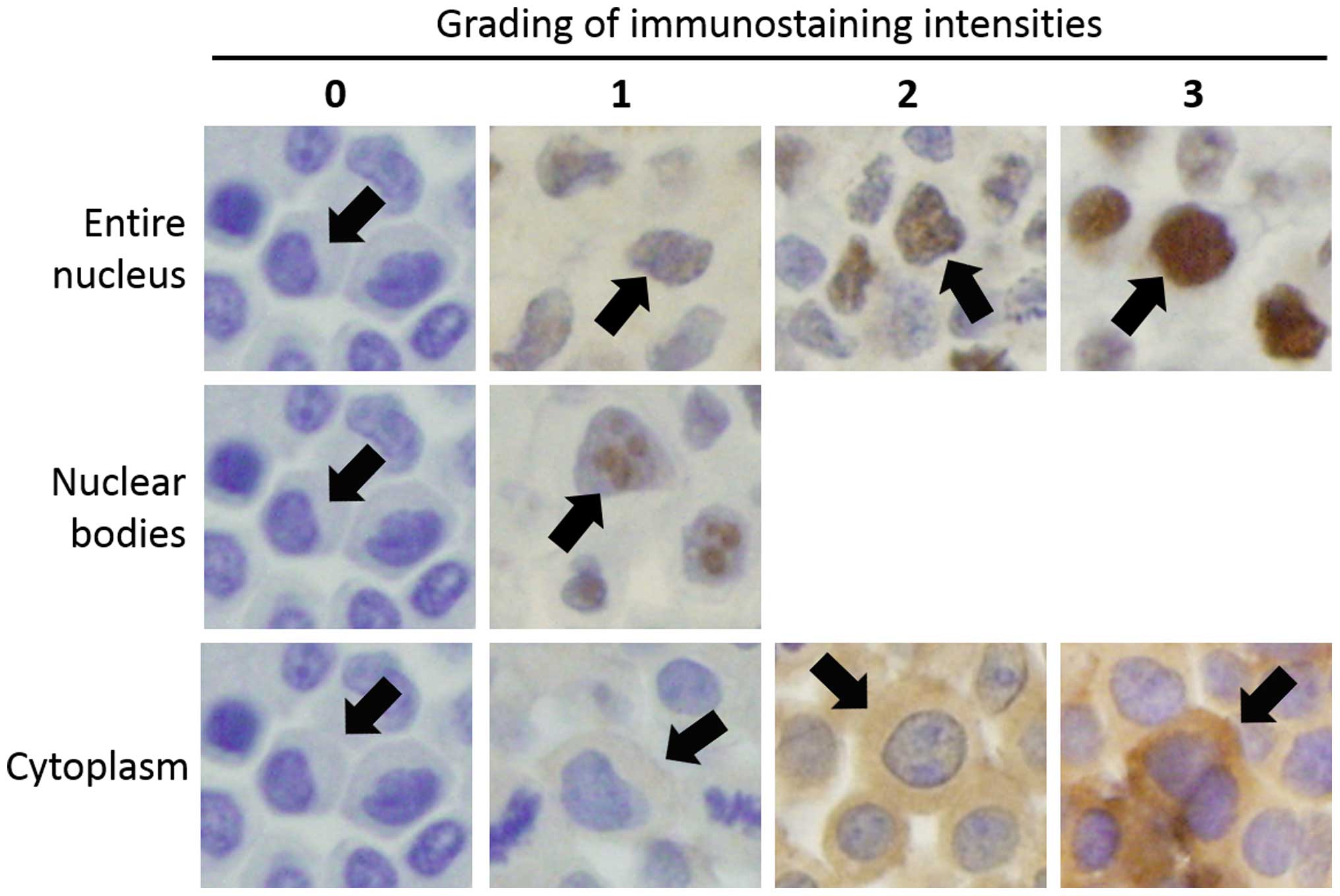
Eosin Aqueous Solution 0.5% (Cytoplasmic Stain) – AbmStore.my

xmlinkhub
xmlinkhub

Fluorescent Cell Stains for Organelles & Cellular Structures | Biotium

Routine Staining in Cytology Laboratory | SpringerLink

Differential detection of cytoplasmic Wilms tumor 1 expression by  immunohistochemistry, western blotting and mRNA quantification
Differential detection of cytoplasmic Wilms tumor 1 expression by immunohistochemistry, western blotting and mRNA quantification